Bioimaging Core Facility

The Bioimaging core facility is specialized in light microscopy. Its goal is to help users from experimental design & acquisitions to image processing (treatment and quantification). In addition to the maintenance of the resources of the platform and the users' training, we are also involved in the organization of workshops about ImageJ and other imaging softwares and in several academic courses. The facility can also help you with data presentation for oral presentation or publication (manipulation of file formats, animations, ...).
PREREQUISITE: If you want to use the Bioimaging core facility for the first time (microscopes and/or computers), please SUBMIT THE REGISTRATION FORM AVAILABLE ON LINE (HERE).
The Bioimaging core facility follows the Directive (in French) newly edited by the Rectorate to optimize the governance, operational procedures and compliance of the UNIGE's Scientific Platforms.
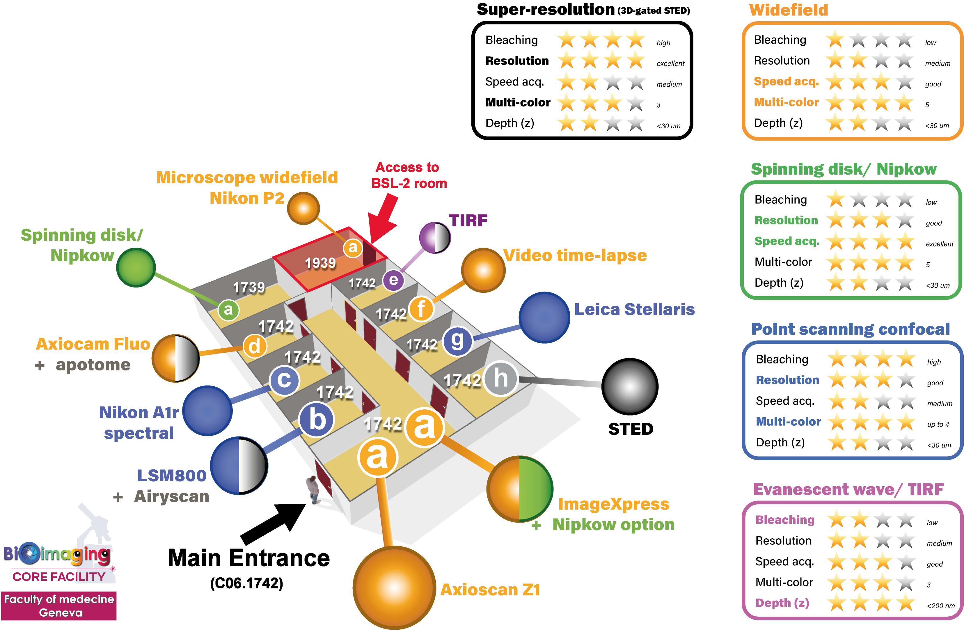
Our bioimaging facility hosts state-of-the-art equipment with:
- 17 imaging systems (1 STED nanoscope, 1 spinning disk confocal microscope (Nipkow), 5 confocal laser scanning microscopes (CLSM), 2 slide scanners and 8 widefield fluorescence microscopes including 1 TIRF evanescent wave microscope, 1 laser microdissector and 1 plate scanner/ImageXpress system) that provide a wide range of applications such as cell membrane visualization, laser microdissection/isolation, colocalization analysis, F-techniques (FRAP, FRET), multi-dimensional time-lapse recordings, intravital imaging and high-throughput imaging in ROOM C06.1742.a.
- 7 PC workstations for image processing and analysis (Imaris, Amira, Huygens, metaXpress, MetaMorph, ZEN Lite, NIS, LAS X FLIM, Fiji, Matlab, QuPath, etc...). These PC are located just in front the microscope in ROOM C06.1542.a.

The LMD is installed in ROOM D06.1554.a.
The Olympus VS120 is installed in ROOM C06.1533.a.


