3D organoids to better understand fatty liver disease
The number of people suffering from fatty liver disease and its complications is increasing worldwide, driven by sedentary lifestyles and hypercaloric diets. The lack of efficacy of existing treatments is prompting a search for new therapeutic options, which often requires the use of mouse models due to the complexity of the disease.
The power of organoids…
Researchers in the laboratory of Obesity, liver metabolic disorders & cancer took a different and innovative approach to study this complex disease. They created 3D liver organoids from human embryonic stem cells which mimic the interactions between the different types of cells. Organoids are a more accurate representation of the liver than traditional 2D cell cultures. They can be produced very quickly compared to mouse models and are also highly reliable. Overall, organoids are a valuable addition to the toolkit for identifying potential therapeutic targets that can help reduce the number of animals used in research.
… to discover new therapeutic targets for fatty liver disease
Using liver organoids that mimic fatty liver disease, the research team discovered that a tinny non-coding nucleic acid, microRNA 149-5p, plays a critical role in the development and progression of the disease. In their recent publication in the Journal of Hepatology Reports, they describe that an overexpression of microRNA 149-5p changes the energy status of cells and promotes the accumulation of fat (as shown in the Figure below) by modulating a large network of genes. Finding a way to inhibit this microRNA, and consequently the entire gene network that it targets in the liver of patients should help to refrain the progression of the disease. These findings not only provide a potential new therapeutic target, but also demonstrate the interest of organoids for the study of fatty liver disease and its complications.
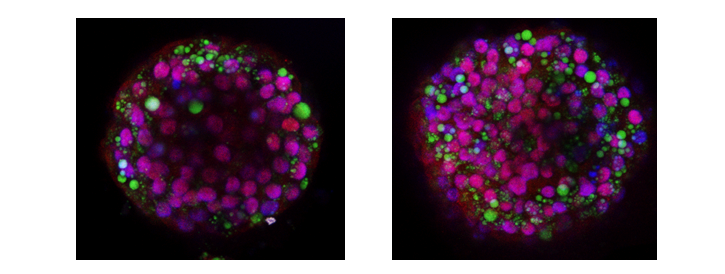
The overexpression of the tinny non-coding nucleic acid microRNA 149-5p promotes the accumulation of fat in liver organoids (green dots, right panel) in comparison to healthy organoids (left panel). Adapted from Figure 4 in Correia de Sousa et al. 2024.