Golgi elements (dictyosomes and vesicles) imaged by cryofracture in maïs and spinach cells.

On the left transmission electron micrograph illustrating a Golgi in a fractured Zea mays root. It consists of stacks of flattened structures forming a dictysome (D) with vesicles at their extremities (red arrows) whose surface is covered of proteins.
Gx: 60.000
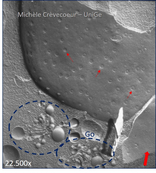
On the right part of a replica in a fractured shoot meristem of spinach. It illustrates the proximity of the nucleus and the Golgi. The dotted blue circle shows the Golgi with stacks of cisternae and vesicles. Red arrows: nuclear pores.
The red arrow at the bottom right indicates the direction of the shading