Publication 73
- “Site-Dependent Excited-State Dynamics of a Fluorescent Probe Bound to Avidin and Streptavidin”
A. Fürstenberg, O. Kel, J. Gradinaru, T.R. Ward, D. Emery, G. Bollot, J. Mareda, E. Vauthey
ChemPhysChem 2009, 10, 1517-1532.
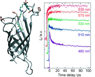
Sensing protein environment: The femtosecond fluorescence dynamics of a molecular probe attached to avidin and streptavidin (see figure) depends markedly on the location of the probe and on the protein. These differences reflect not only the protein primary and tertiary structures but also the effect of the surrounding water molecules.
The excited-state dynamics of biotin–spacer–Lucifer-Yellow (LY) constructs bound to avidin (Avi) and streptavidin (Sav) was investigated using femtosecond spectroscopy. Two different locations in the proteins, identified by molecular dynamics simulations of Sav, namely the entrance of the binding pocket and the protein surface, were probed by varying the length of the spacer. A reduction of the excited-state lifetime, stronger in Sav than in Avi, was observed with the long spacer construct. Transient absorption measurements show that this effect originates from an electron transfer quenching of LY, most probably by a nearby tryptophan residue. The local environment of the LY chromophore could be probed by measuring the time-dependent polarisation anisotropy and Stokes shift of the fluorescence. Substantial differences in both dynamics were observed. The fluorescence anisotropy decays analysed by using the wobbling-in-a-cone model reveal a much more constrained environment of the chromophore with the short spacer. Moreover, the dynamic Stokes shift is multiphasic in all cases, with a ~1 ps component that can be ascribed to diffusive motion of bulk-like water molecules, and with slower components with time constants varying not only with the spacer, but with the protein as well. These slow components, which depend strongly on the local environment of the probe, are ascribed to the motion of the hydration layer coupled to the conformational dynamics of the protein.
DOI : 10.1002/cphc.200900132
archive ouverte unige:3554